Sickle Cell Retinopathy
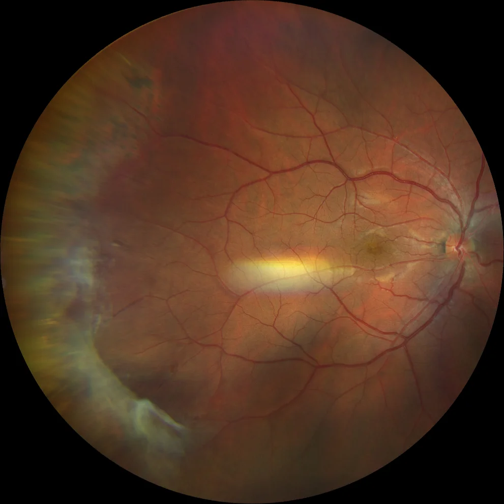
A and B. Color retinography (Clarus 500, Carl Zeiss Meditec ASG, Jena, Germany) of the right and left eyes of a patient with sickle cell retinopathy, showing fans of fibrous neovascular tangles in the temporal periphery of both eyes after treatment with photocoagulation and anti-VEGF.
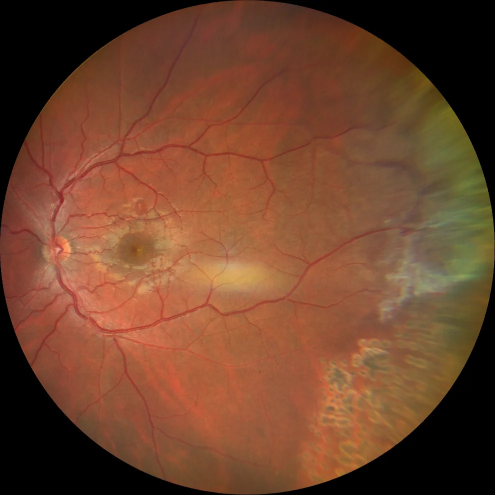
A and B. Color retinography (Clarus 500, Carl Zeiss Meditec ASG, Jena, Germany) of the right and left eyes of a patient with sickle cell retinopathy, showing fans of fibrous neovascular tangles in the temporal periphery of both eyes after treatment with photocoagulation and anti-VEGF.
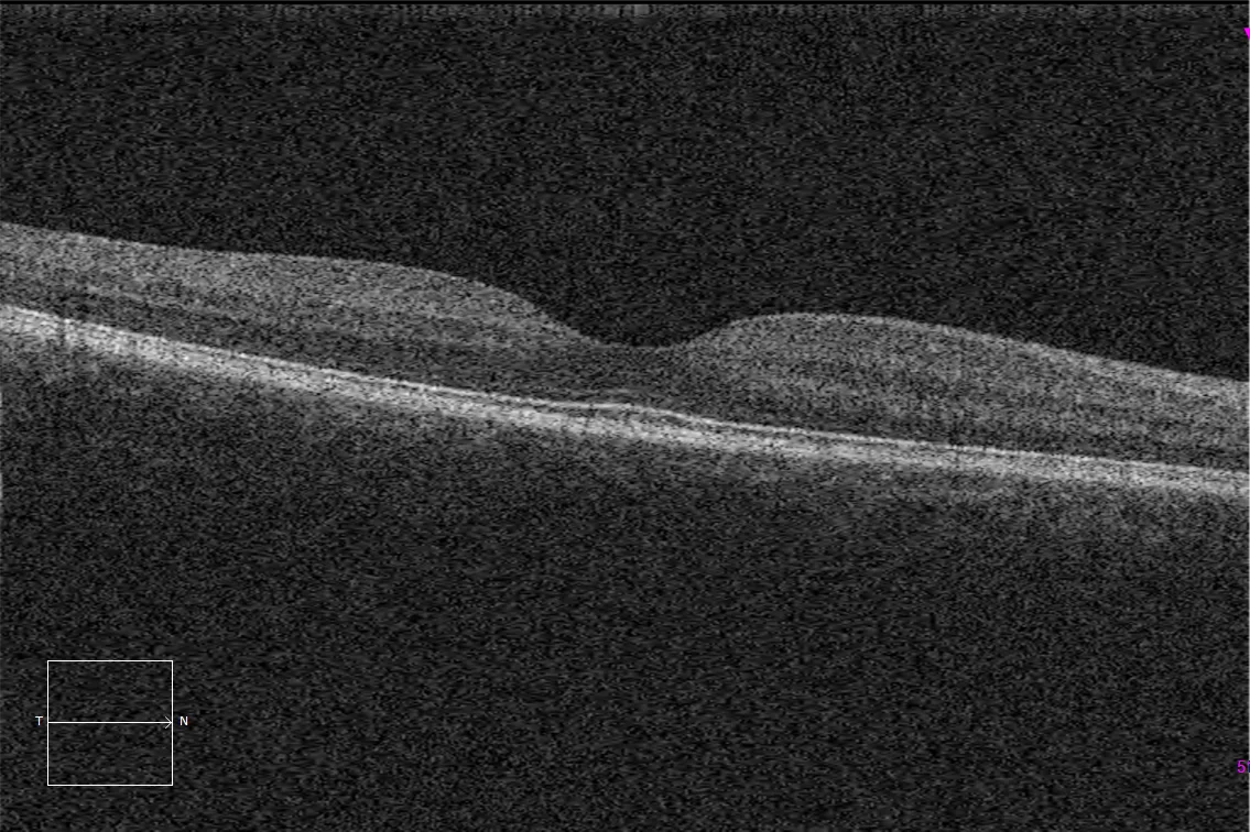
C and D. Macular OCT (Cirrus 5000, Carl Zeiss Meditec ASG, Jena, Germany) of the right and left eyes, without pathological alterations.
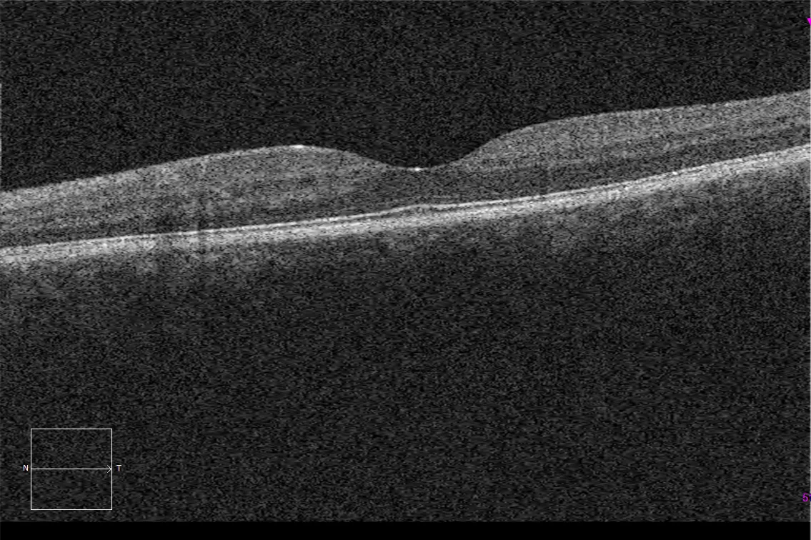
C and D. Macular OCT (Cirrus 5000, Carl Zeiss Meditec ASG, Jena, Germany) of the right and left eyes, without pathological alterations.
Description
Sickle cell retinopathy can be present in patients with sickle cell disease, an autosomal recessive hemoglobinopathy caused by a mutation in the gene that encodes the beta chain of hemoglobin.
It progresses with an initial non-proliferative phase in which areas of peripheral ischemia and hemorrhages with a characteristic salmon-colored appearance develop. Visual loss may occur due to macular ischemia.
This chronic ischemia can lead to the formation of new blood vessels in the proliferative phase.