Soft drusen and serous PED
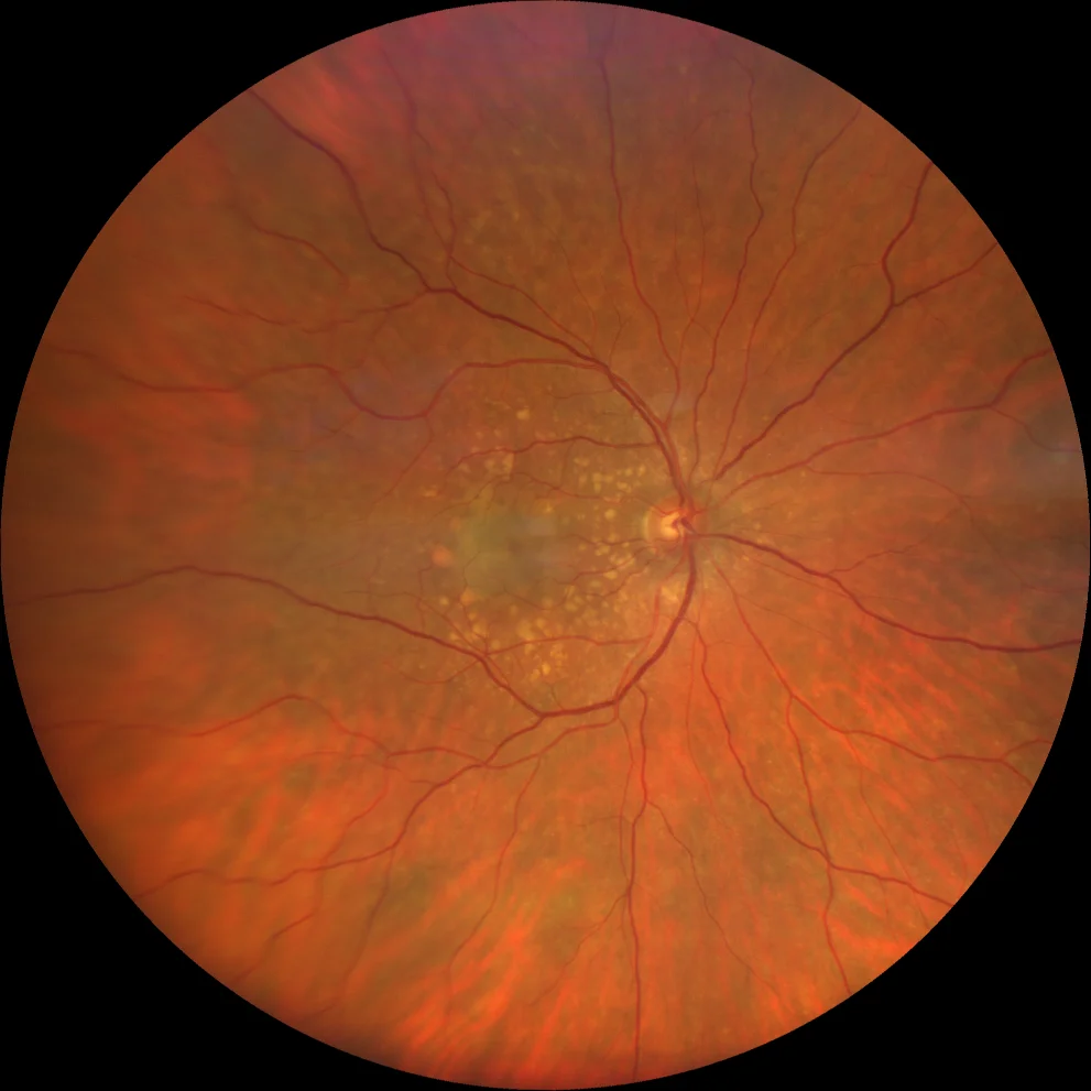
A: Soft drusen appear as yellowish lesions with smooth edges and a tendency to coalesce. A poorly defined greyish area can be seen in the central region.
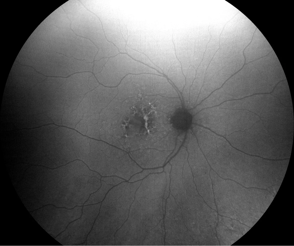
B (green AF): hyperAF areas alternate with hypoAF areas, reflecting the alteration of the EPR.
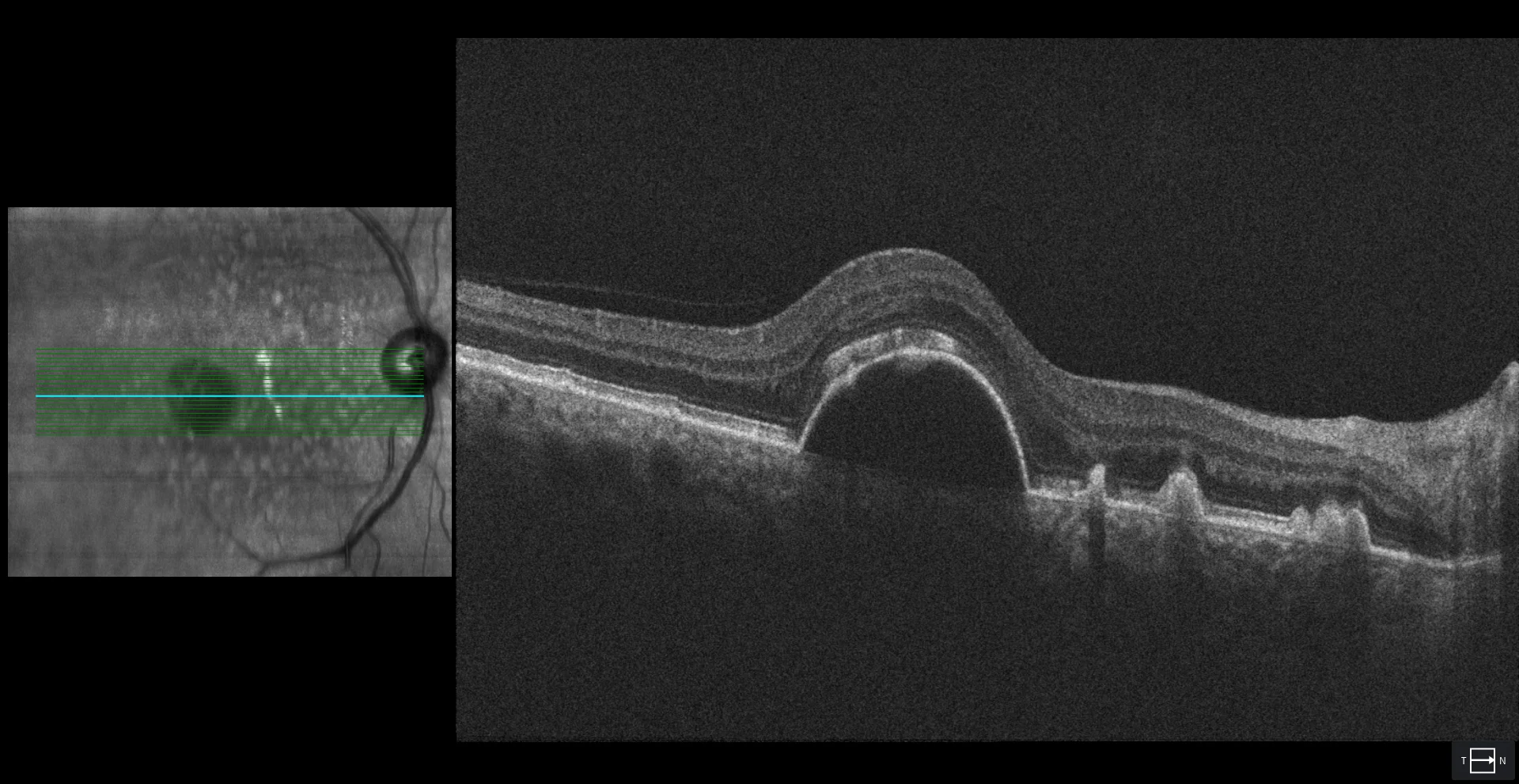
C: Soft drusen are dome-shaped elevations of the RPE with heterogeneous content (hyper-, iso- or hyporeflective). Serous PED is a large, rounded elevation of the RPE with hyporeflective content. It corresponds to the poorly defined grayish area seen on the retinography (A). It presents an overlying subretinal hyperreflective material that corresponds to vitelliform material that forms at the apex of the serous PED. No lesions suggestive of type 1 VAP are seen on OCT.
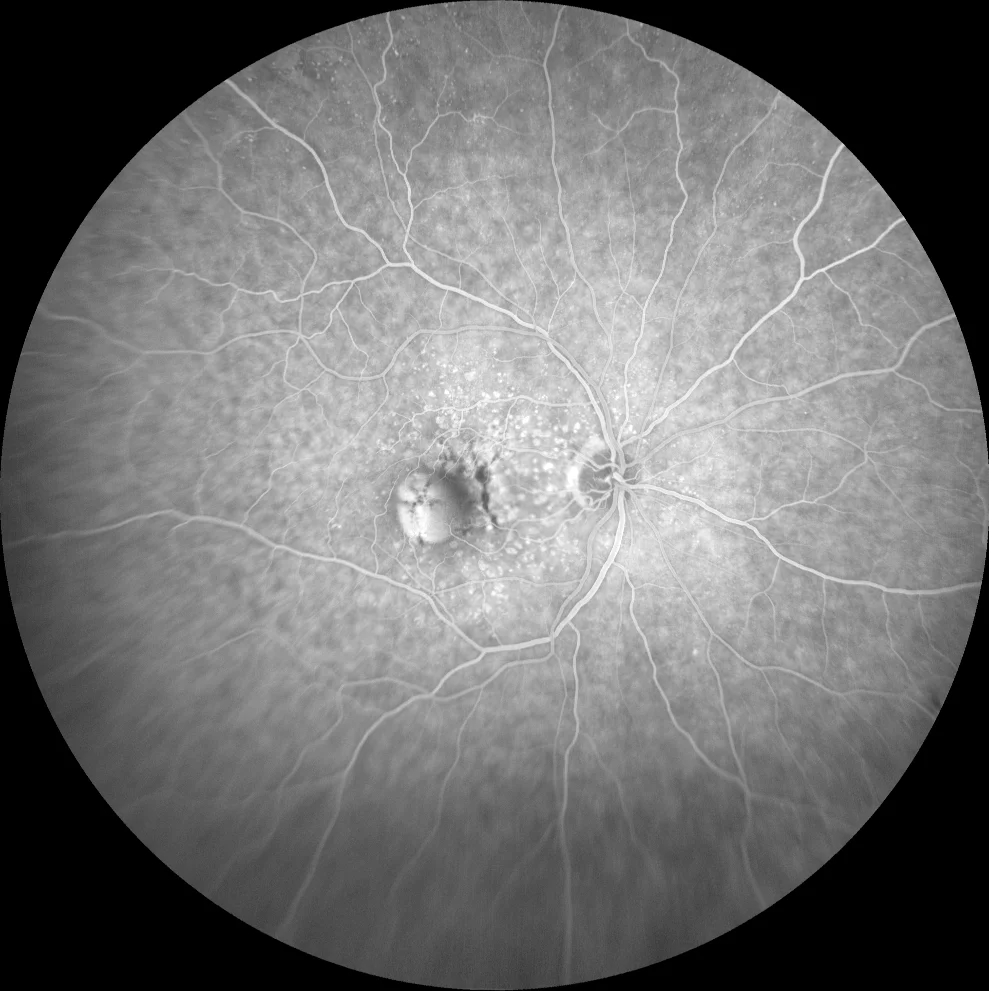
D1, D2: soft drusen show early hyperF due to the window effect (due to atrophy of the RPE at the apex) (D1) which does not produce leakage in later times (D2). The serous PED shows hyperF from early times and remains in late times due to fluorescein accumulation (pooling) in its interior. No lesions suggestive of occult neovascularization are observed.
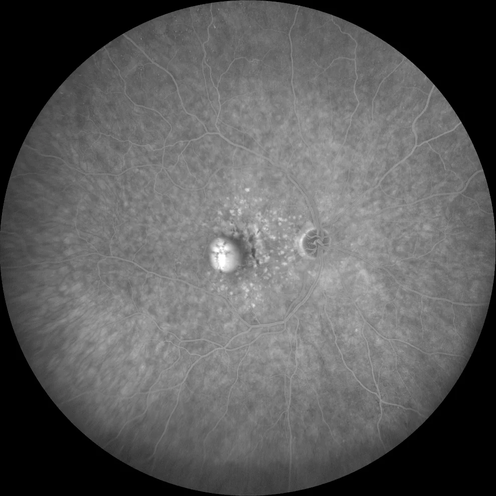
D1, D2: soft drusen show early hyperF due to the window effect (due to atrophy of the RPE at the apex) (D1) which does not produce leakage in later times (D2). The serous PED shows hyperF from early times and remains in late times due to fluorescein accumulation (pooling) in its interior. No lesions suggestive of occult neovascularization are observed.
Description
73-year-old woman with vision loss and metamorphopsia in the right eye of 1 month’s duration
AVMC OD 20/50 OI 20/20
Soft drusen in both eyes. Serous PED without associated neovascularization in the right eye.