Solitary congenital hypertrophy of the retinal pigment epithelium (CHPRE)

Color retinography: solitary HCEPR: pigmented lesion with lacunae inside followed for 25 years

WF color retinography: solitary HCEPR and temporal chorioretinal folds
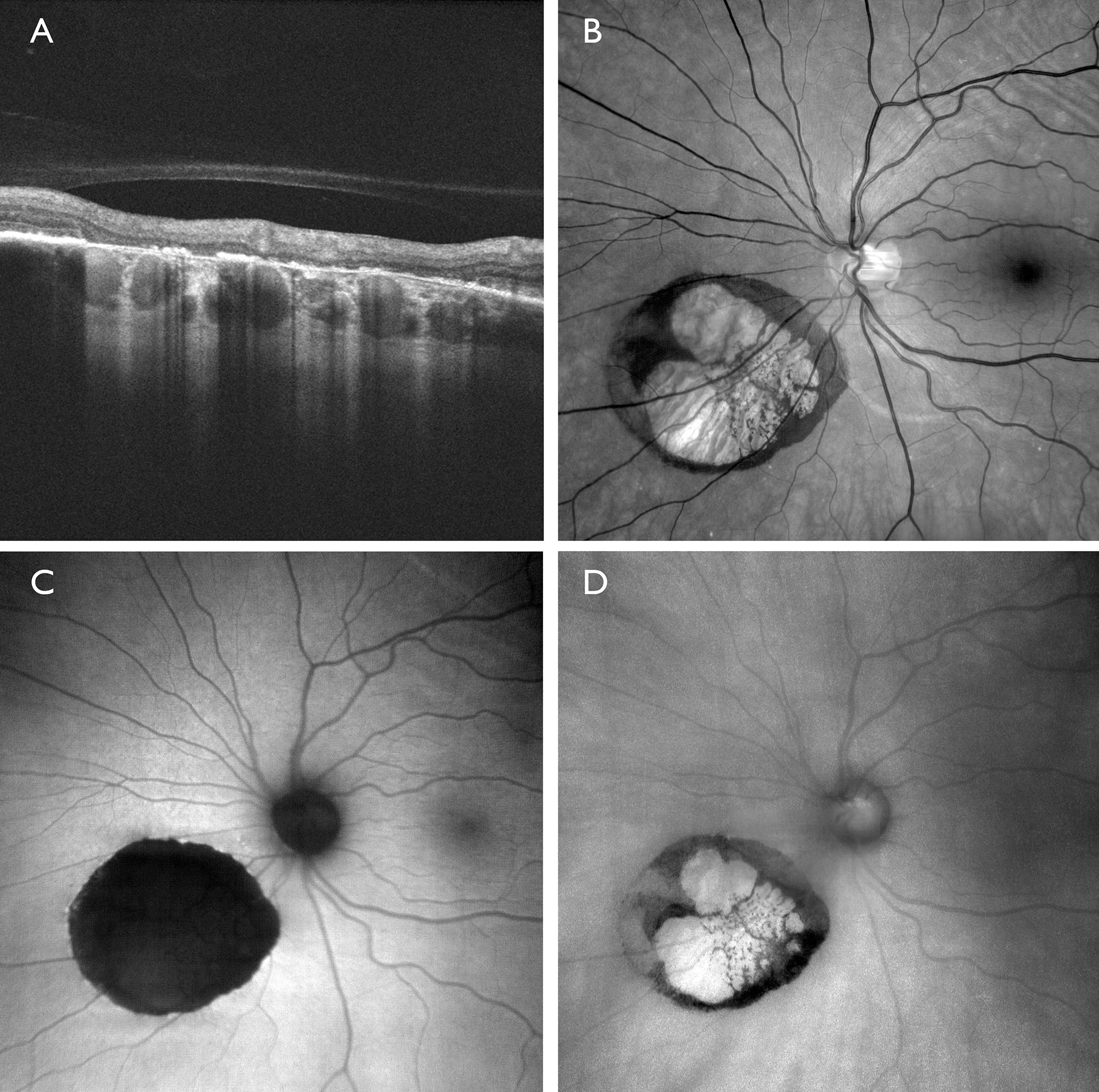
Multimodal image: Solitary HCEPR - OCT (A): EPR with increased signal at the edges, with thinning of the neurosensory retina and lacunae with increased signal passage inside. - Green filter (B): visualization of the lesion by location in the EPR - Green autofluorescence (C): intense and well-defined hypoautofluorescence - Infrared (D): Pigmented area is highlighted
Description
Solitary HCEPR is a single, highly pigmented, flat lesion with well-defined borders, usually located in the mid-periphery. It may be surrounded by a halo of pigmentation and contain clear lacunae in 43% of cases. It is usually asymptomatic, may experience slow growth, and is not associated with systemic pathology.