Syphilitic posterior placoid chorioretinitis
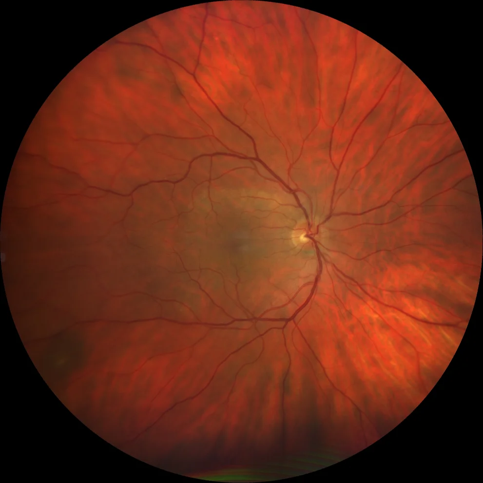
A. Color retinography of the right eye (Clarus 500, Carl Zeiss Meditec ASG, Jena, Germany) showing a yellowish placoid lesion covering almost the entire macula.
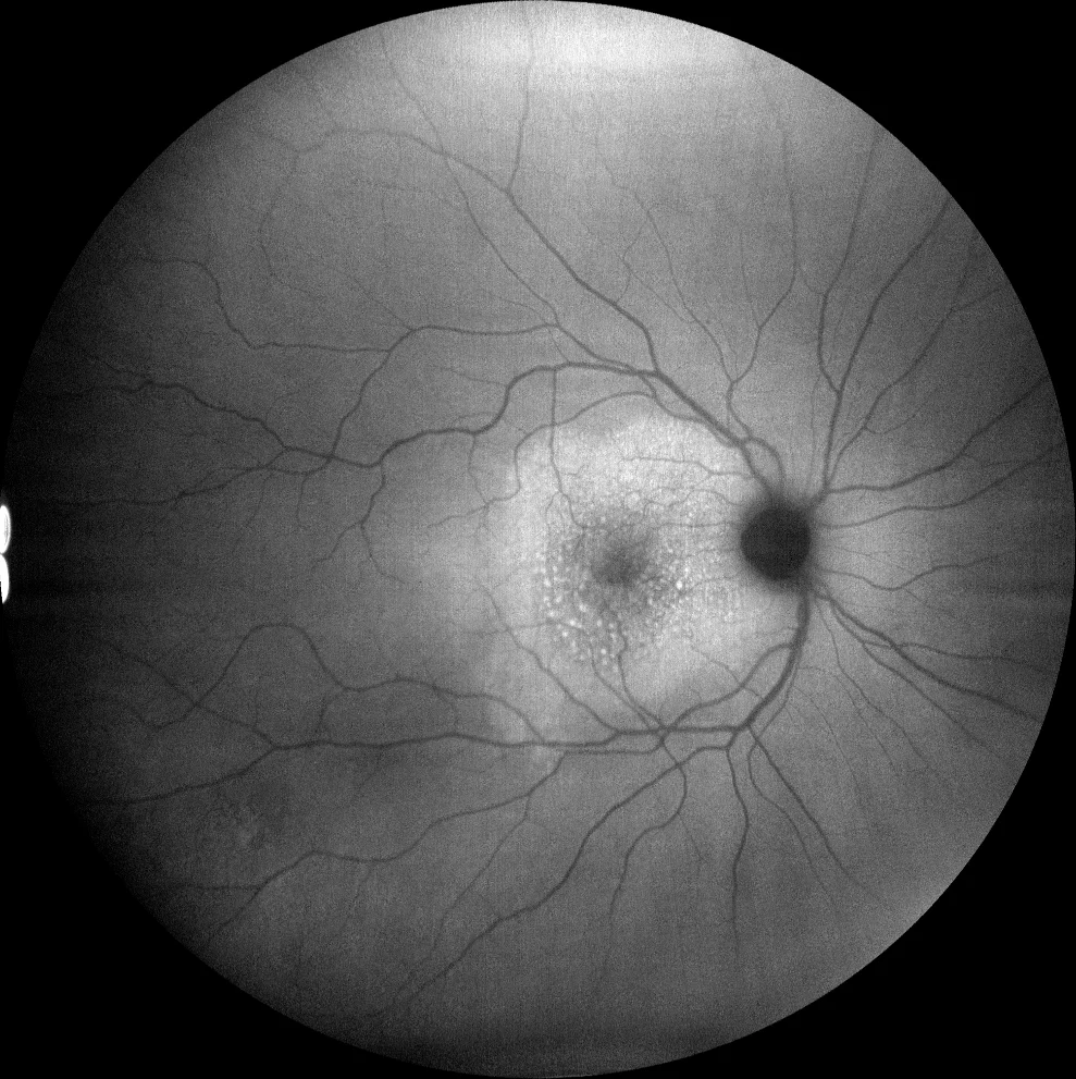
B. Autofluorescence image (Clarus 500, Carl Zeiss Meditec ASG, Jena, Germany) showing diffuse hyperautofluorescence in the placoid lesion with even more autofluorescent granules.
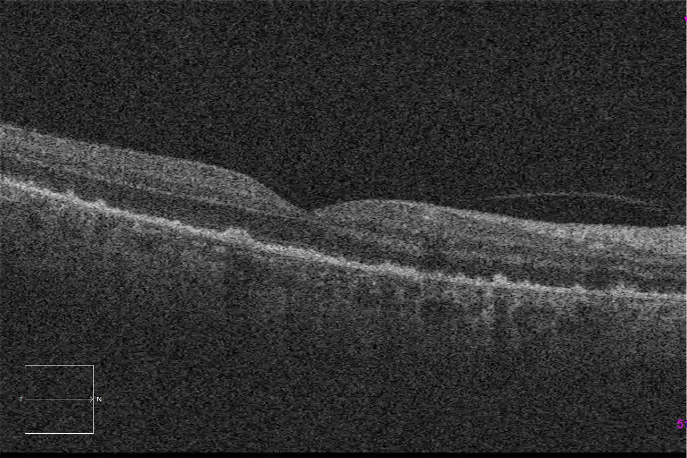
C. Macular optical coherence tomography (Cirrus 5000, Carl Zeiss Meditec ASG, Jena, Germany) on the placoid lesion, showing a segmental loss of the ellipsoid and external limiting membrane with preservation of the latter at the subfoveal level, together with hyperrefringent accumulations at the level of the pigmented epithelium, characteristic of this nosological entity.
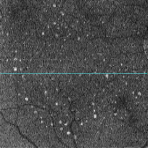
D. En face OCT image at the level of the ellipsoids (Cirrus 5000, Carl Zeiss Meditec ASG, Jena, Germany), showing the distribution of hyperrefringent clusters, which correspond to the hyperautofluorescent granules of the autofluorescence.
Description
Syphilis is a sexually transmitted infection caused by the spirochete Treponema pallidum . Posterior placoid chorioretinitis is a rare but highly characteristic clinical manifestation of ocular syphilis. Neurosyphilis and HIV co-infection should be ruled out in all patients with syphilitic posterior placoid chorioretinitis. Early treatment with intravenous penicillin is often effective and may help improve visual acuity in these patients.