Torpedo maculopathy associated with choroidal neovascularization (CNV)
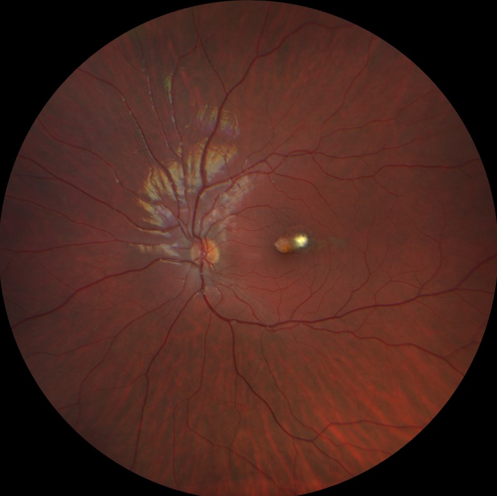
Color retinography (Clarus 500, Carl Zeiss Meditec ASG, Jena, Germany) of the left eye showing a single depigmented temporal macular lesion with a whitish area at the tail of the lesion.
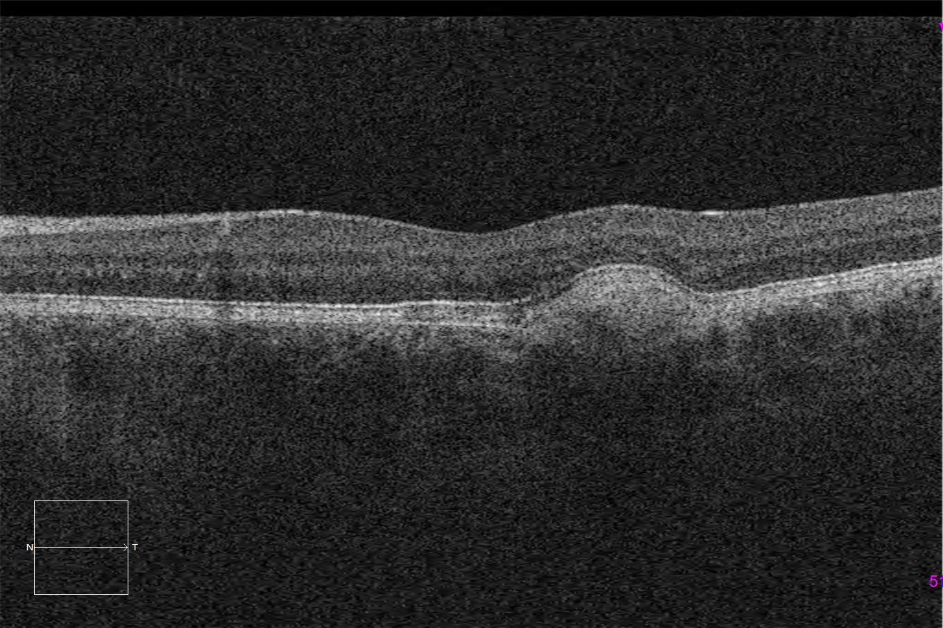
Macular optical coherence tomography (Cirrus 5000, Carl Zeiss Meditec ASG, Jena, Germany) of the lesion in the left eye, showing a RPE defect in the most nasal area, along with an elevation composed of subretinal hyperintense material in the most temporal part. The outer retinal layers appear preserved.
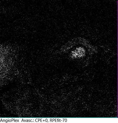
OCT- angiography (Cirrus 5000, Carl Zeiss Meditec ASG, Jena, Germany) of the avascular layer of the left eye, showing choroidal neuvavascularization in the most temporal part of the lesion.
Description
Torpedo maculopathy is a developmental macular disorder. It presents as an elongated, unilateral scar, mostly horizontal, with a whitish “head.” Bilateral presentation has been described, as well as oblique torpedo defects that do not directly affect the foveal area. These scars result from a developmental defect of the retinal pigment epithelium (RPE) in the median raphe. They do not usually have functional repercussions since the outer layers are typically preserved, but they are sometimes associated with neurovascular bundles (NVBs), as in this case, and therefore require follow-up.
The diagnosis is usually made at early ages due to the presence of a macular scar that is difficult to classify.