Type 1 macular neovascularization with aneurysmal dilatations
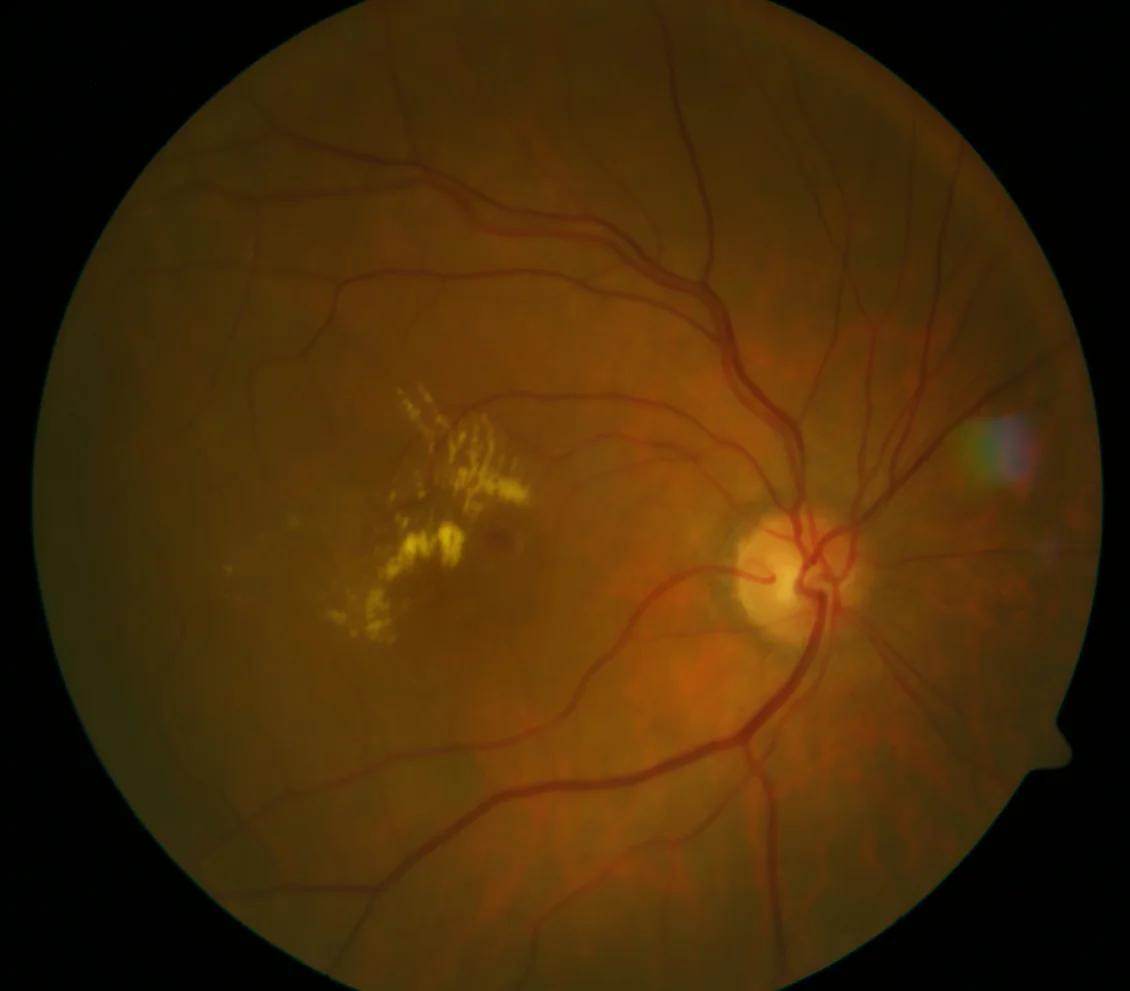
A: Multiple hard exudates in the temporal macular area accompanied by a rounded reddish-brown lesion close to the fovea
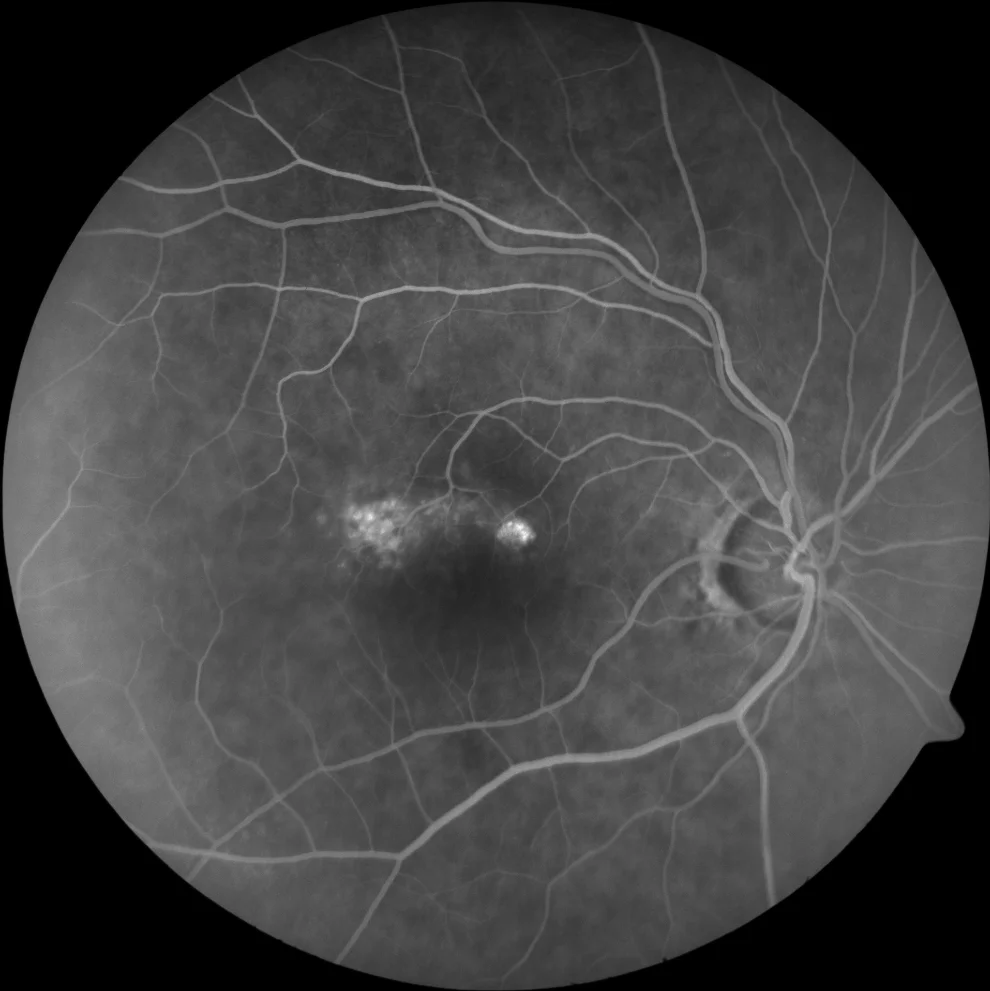
Discrete, ill-defined hyperfluorescence in early stages (B1) that increases slightly in late stages (B2)
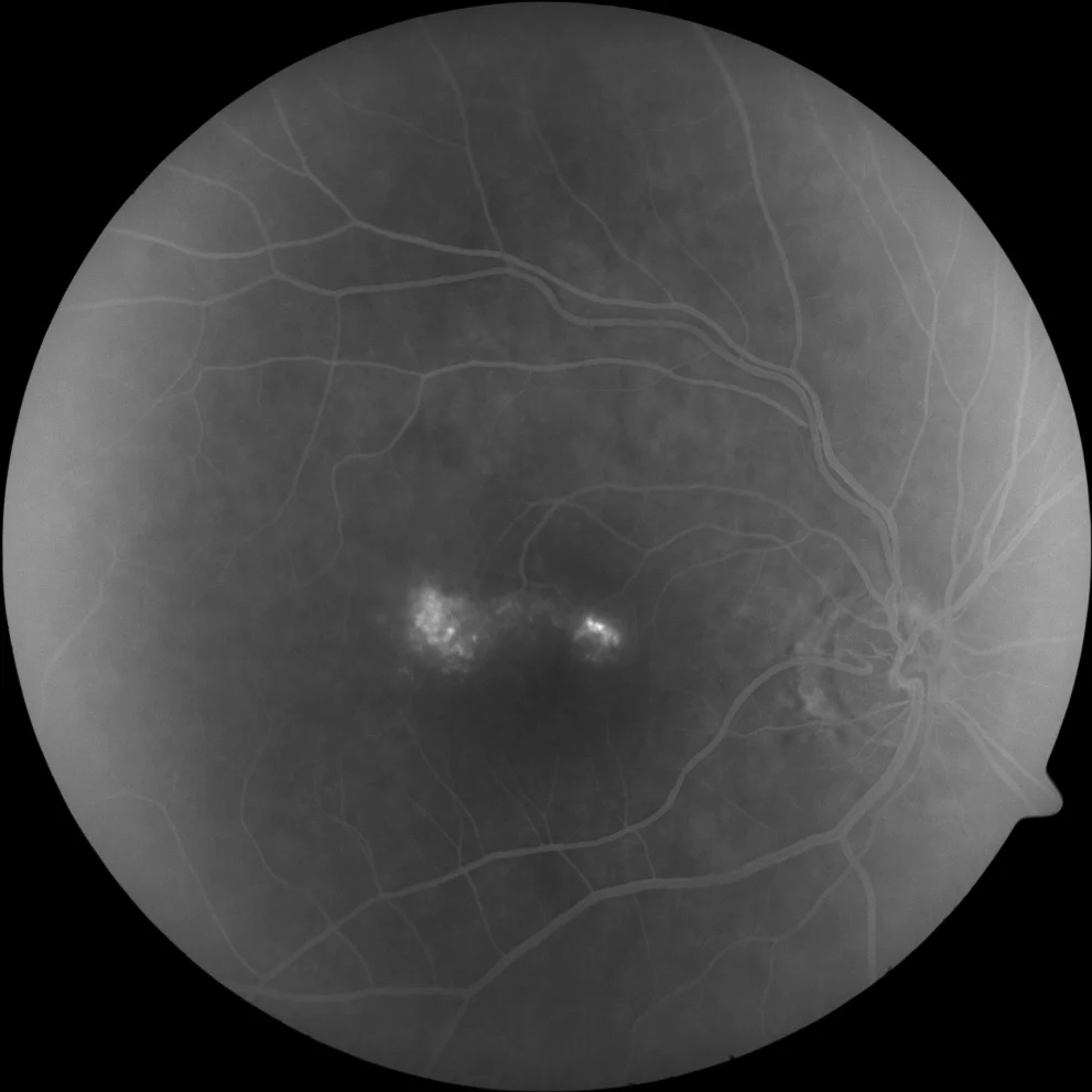
Discrete, ill-defined hyperfluorescence in early stages (B1) that increases slightly in late stages (B2)
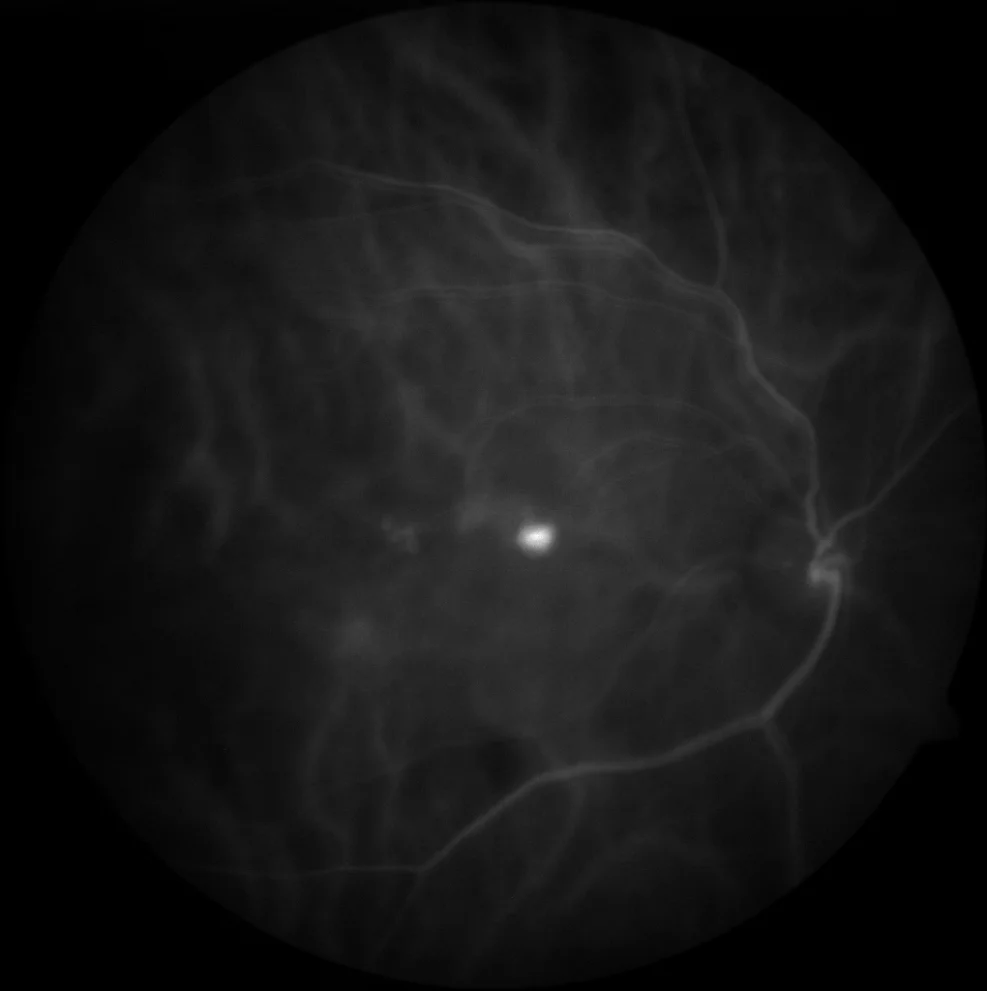
The rounded lesion shows very intense hyperfluorescence in early stages that corresponds to an aneurysmal dilatation and is accompanied by a slightly hyperfluorescent lesion towards the temporal (type 1 NVM, previously called branching vascular network)
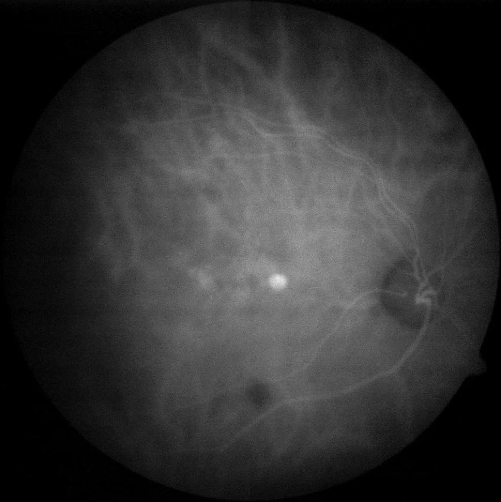
The hyperfluorescence of both components is maintained at late times
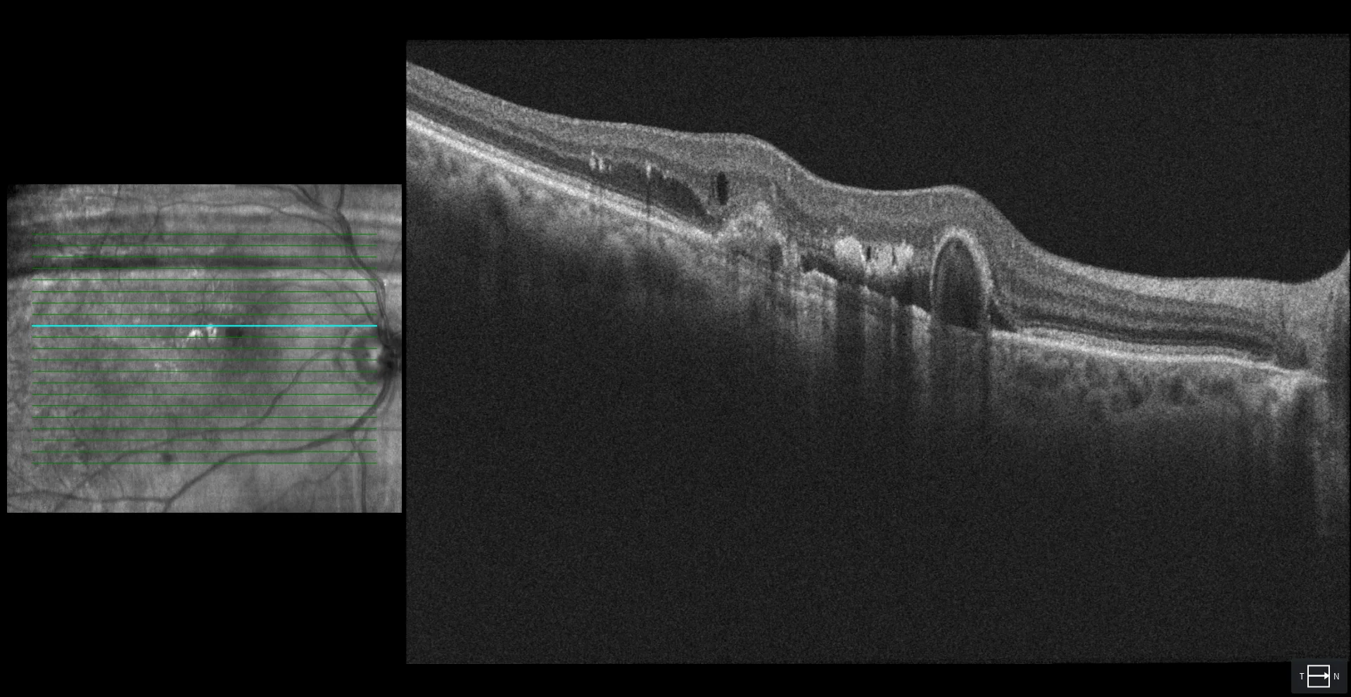
Aneurysmal dilatation is seen as an omega-shaped elevation of the RPE. Adjacent to it is a flat irregular elevation of the RPE (SIRE) that corresponds to type 1 NVM. In addition, sub- and intraretinal fluid and confluent hyperreflective foci that correspond to hard exudates are observed.
Description
75-year-old woman who comes due to loss of vision in her right eye.
VA in OD is 20/80. It is phakic.
The fundus reveals multiple hard exudates in the temporal macular region and a reddish-brown lesion near the fovea. Fluorescein angiography, indocyanine green angiography and OCT allow the patient to be diagnosed with type 1 NVM with aneurysmal dilatations.