Sección: High Myopia
Myopia Case 1 Tiled Background
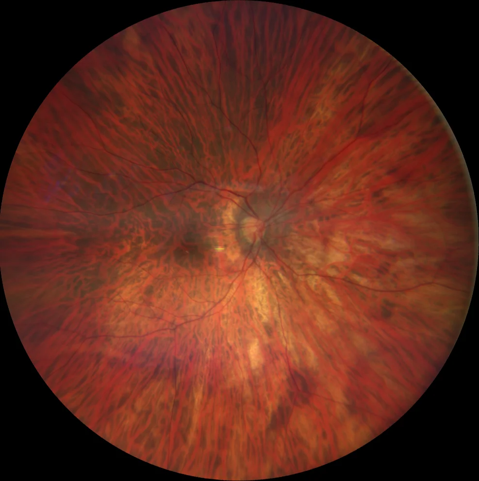
43-year-old woman who attends regular check-ups.
AV OD 20/25 with -18.50.
AV OI 20/25 with -19.00.
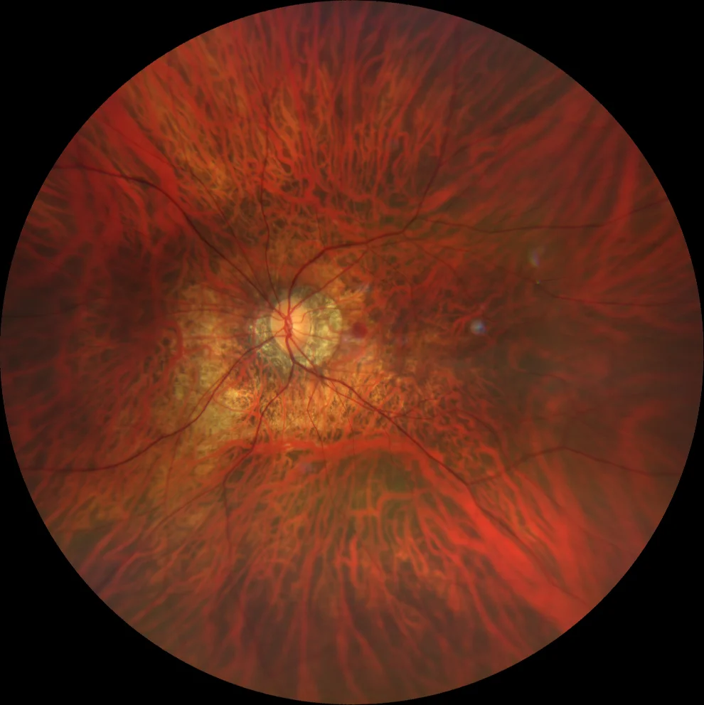
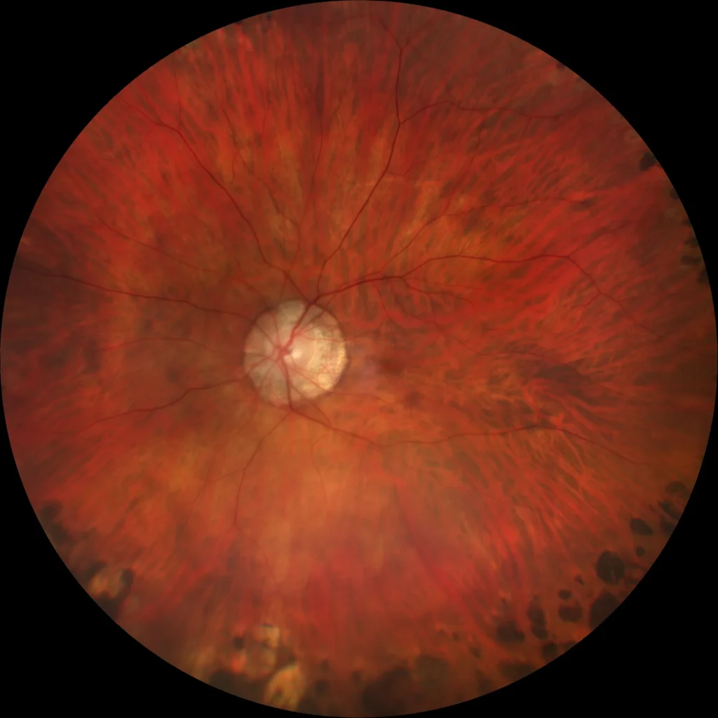
A very tessellated fundus is observed in both ey
Tiled Background 2
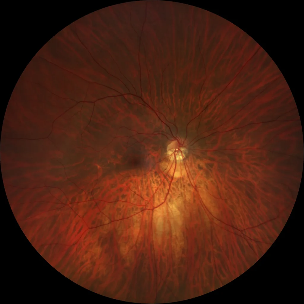
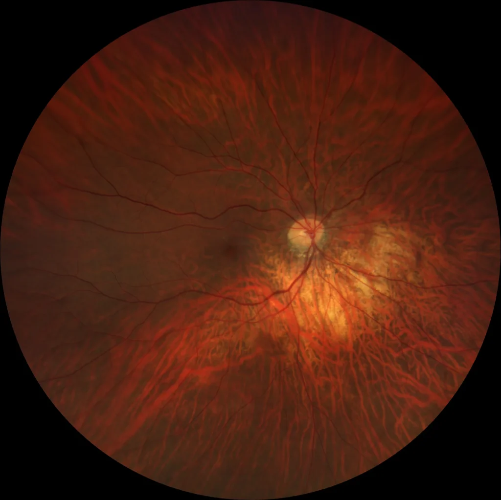
Increased visibility of the large choroidal vessels as a result of pigment loss and attenuation of the retinal pigment epithelium secondary to axial e
Tiled background
Increased visibility of the large choroidal vessels as a result of pigment loss and attenuation of the retinal pigment epithelium secondary to axial e
Myopic retinoschisis
Progressive separation of the retinal layers caused by the existence of a posterior staphyloma that generates an external traction, in combination wit
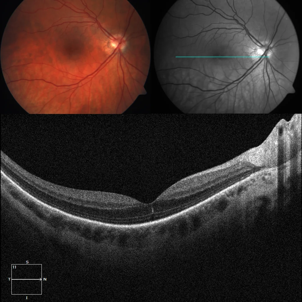
Myopic retinoschisis
Myopic retinoschisis or foveoschisis was described by Takano in 1999. It is included within tractional myopic maculopathy. Myopic foveoschisis may pre
Myopic neovascularization
It is defined as a neovascularization of choroidal origin (neovascular membrane) in the context of pathological myopia. Typically, they tend to be sub

Macula in dome
Gaucher defined it as a convex protrusion within a posterior staphyloma, although it is currently considered a new form of staphyloma. Its pathogenesi
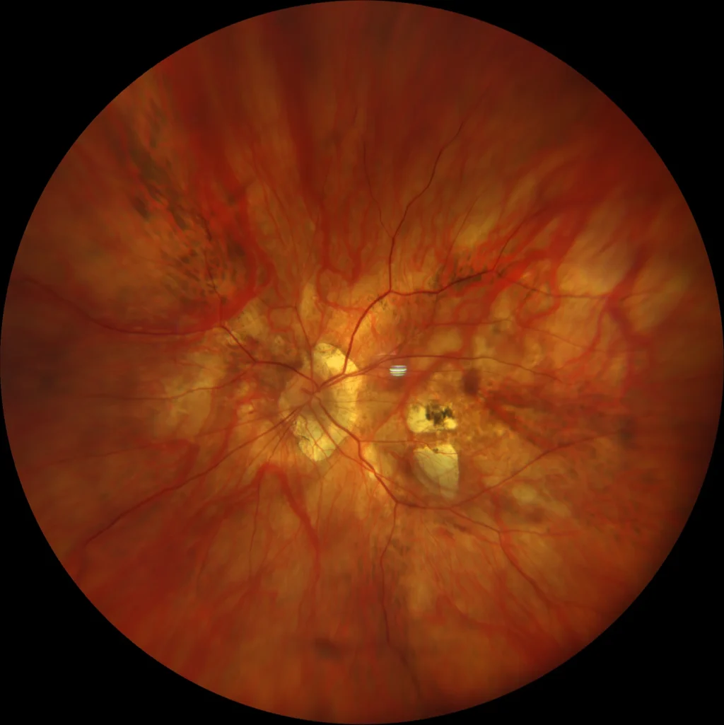
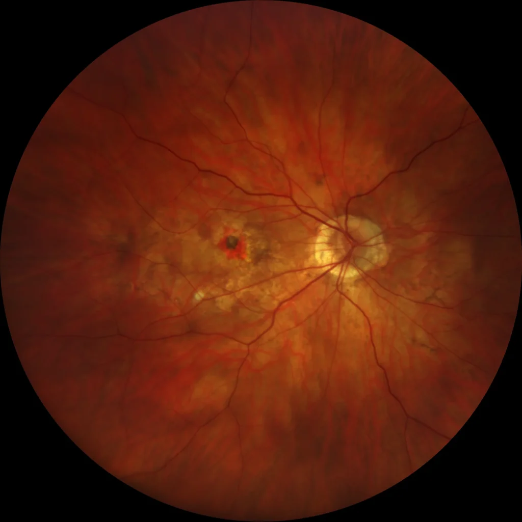
Myopic macular hemorrhage
A 47-year-old male comes to the doctor due to sudden loss of vision in his right eye.
AV OD 20/40 with -9.00 -4.25 x 160º.
Fundus examination revea
Vanishing hemorrhage
Evanescent hemorrhages in myopes are usually subretinal hemorrhages (sometimes intraretinal) that occur during the development or expansion of a lacqu