Sección: Vitreomacular Interface Pathology
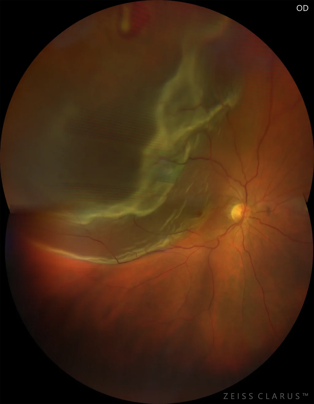
Posterior Vitreous Detachment
Posterior vitreous detachment is the process by which the posterior hyaloid partially or completely separates from the retina. In most cases, it does
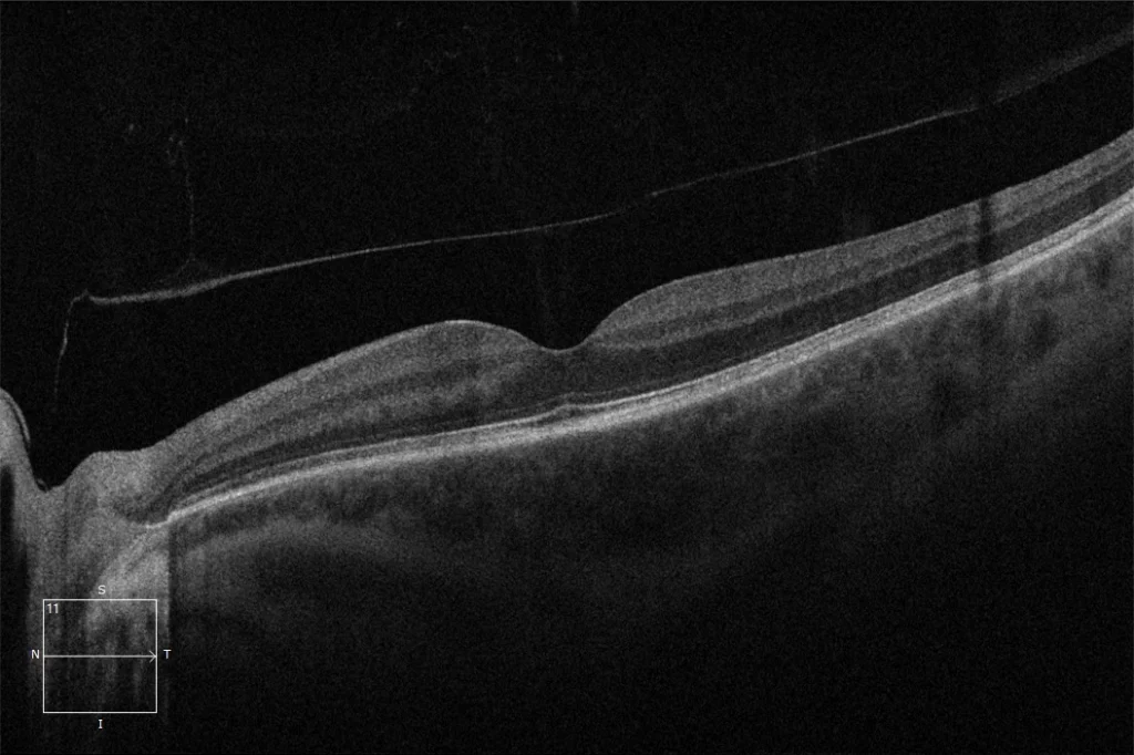
Epiretinal Membrane (ERM) Associated with Macular Pseudohole
The ERM generates a traction force on the macula; when this force is centrifugal, it causes a separation of the retinal layers in the fovea, which is
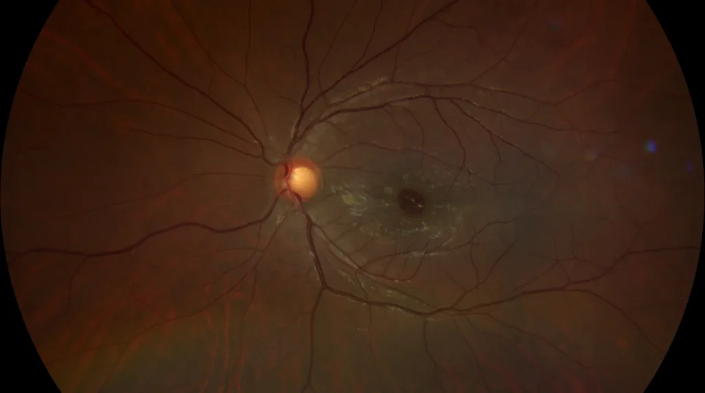
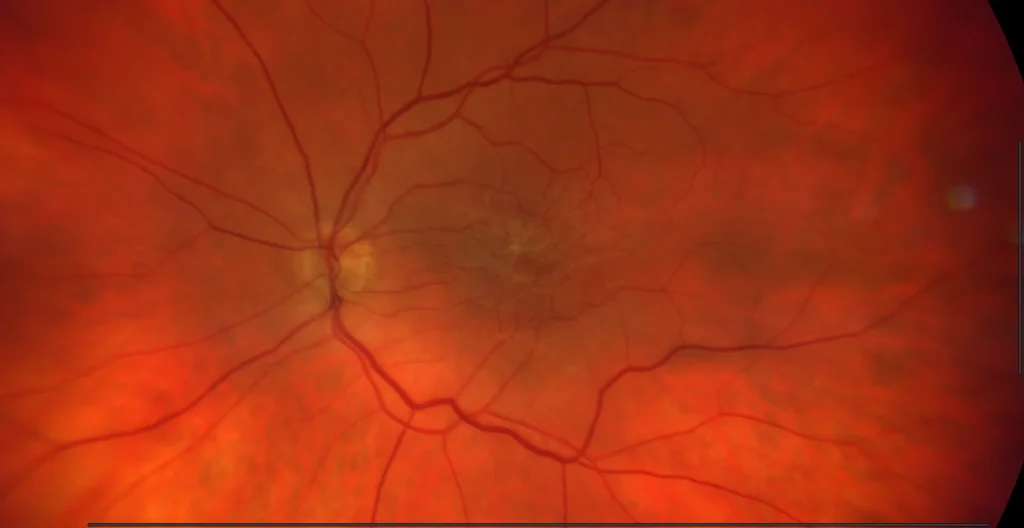
Epiretinal Membrane (ERM)
Epiretinal Membrane (ERM). It is a fibrocellular, avascular, and transparent tissue located on the inner surface of the retina that adheres to and cov
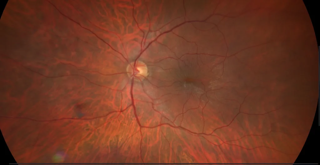
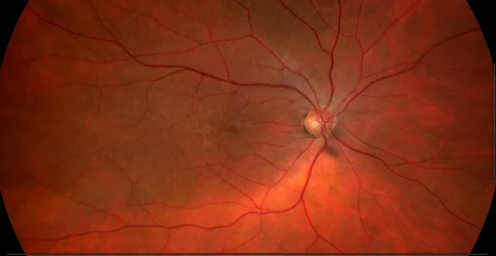
Epiretinal Membrane (ERM)
Epiretinal Membrane (ERM). It is an avascular, transparent fibrocellular tissue located on the internal surface of the retina that adheres to and cove
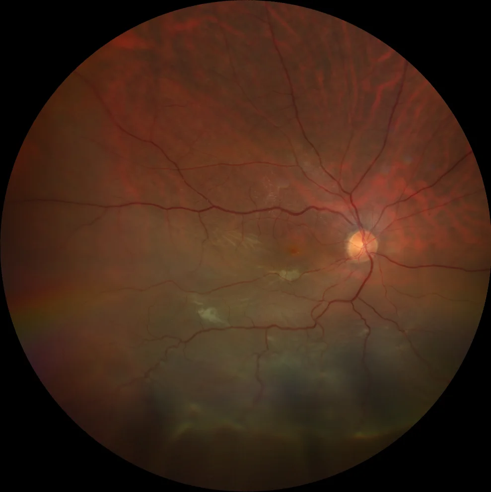
Epiretinal Membrane (ERM)
Epiretinal Membrane (ERM). It is an avascular, transparent fibrocellular tissue located on the internal surface of the retina that adheres to and cove
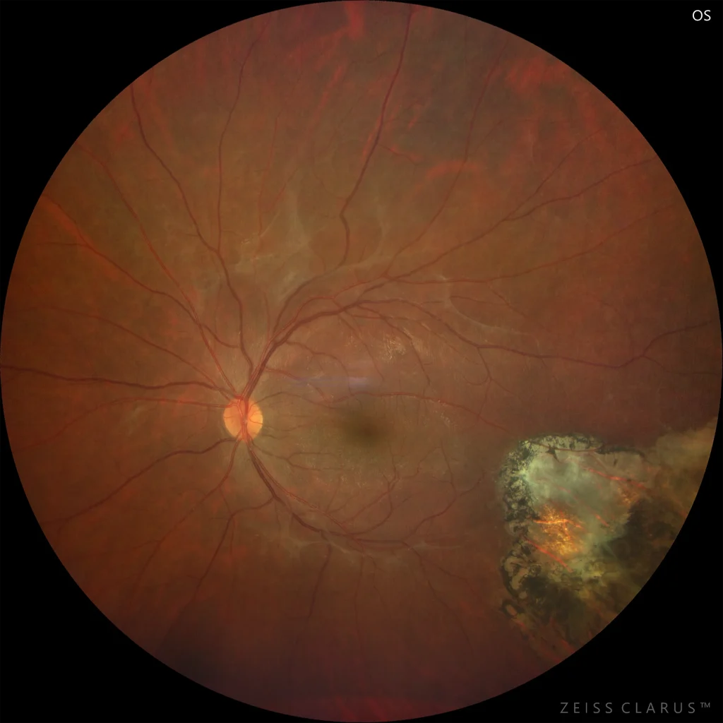
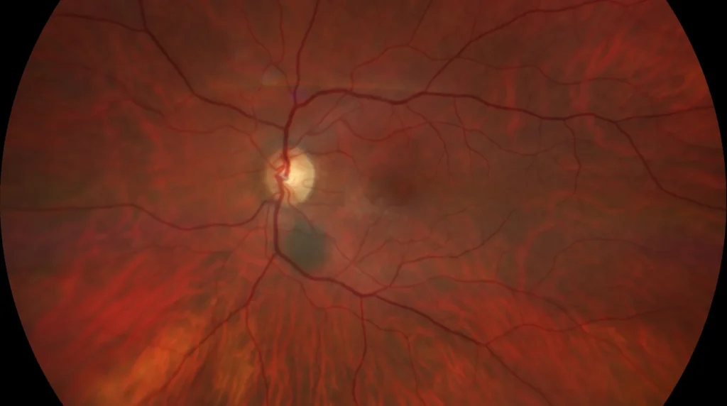
Macular epiretinal membrane in a patient with toxoplasmosis
Toxoplasmosis is the most common cause of infectious retinochoroiditis in humans. Characteristic features include a pigmented retinochoroidal scar, fo
Rhegmatogenous retinal detachment (RRD)
The word “rhegmatogenous” comes from the Greek word rhegma , meaning break. RRDs are caused by the passage of fluid from the vitreous cavity into
Rhegmatogenous retinal detachment
Rhegmatogenous retinal detachment is a process in which, mostly spontaneously, a separation of the neurosensory retina from the retinal pigment epithe