Diffuse Choroidal Atrophy

Color retinography (Clarus500, Zeiss): (A) OD: Diffuse choroidal atrophy affecting the peripapillary area and posterior pole. Peripapillary chorioretinal atrophy. (B) OS: Diffuse choroidal atrophy affecting the peripapillary area and posterior pole with granular pigmentary alteration in the macula. Peripapillary and superior to the macula chorioretinal atrophy plaques.
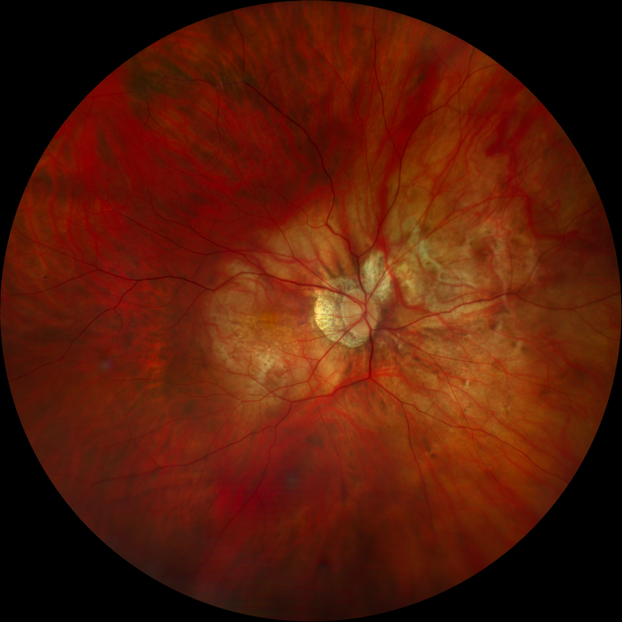
Retinography OD
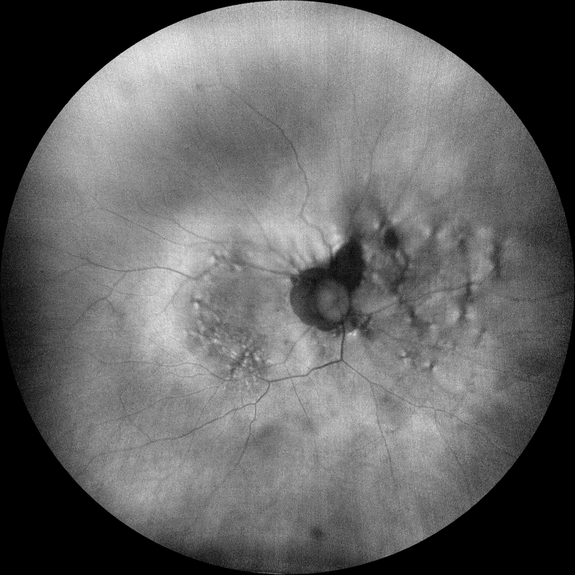
Autofluorescence (Clarus500, Zeiss) OD
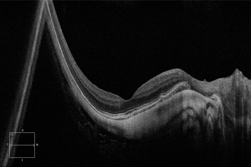
OCT (Cirrus6000, Zeiss) (E) OD

Autofluorescence OS

Retinography OS
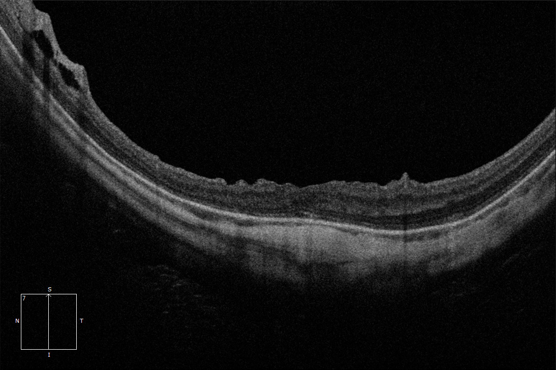
Macular OCT OS
Description
Diffuse Choroidal Atrophy. Corresponds to category 2 of atrophic myopic maculopathy. It presents as a poorly defined yellowish lesion in the posterior pole of high myopes, which is not uniform and may have a granular appearance. It usually begins around the optic nerve and progresses to affect the entire posterior pole. Unlike tessellated fundus, the frequency of this lesion increases with age and axial length, being more prevalent over 40 years (30-52.9%) and above 28.5 mm of axial length (62.8%). OCT can show marked choroidal thinning; however, the retina and RPE are usually preserved, which could explain relatively preserved vision in these patients.