geographic atrophy
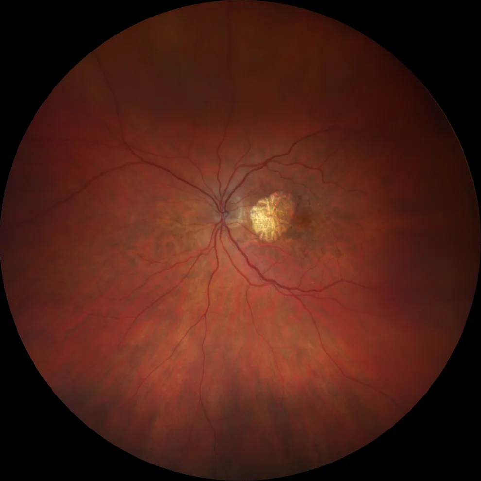
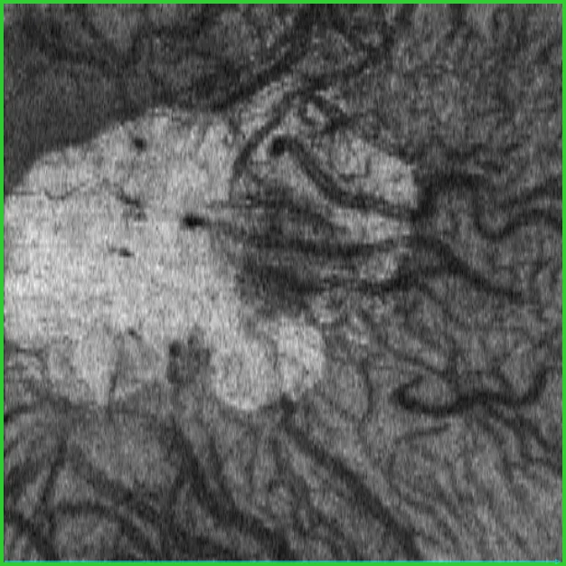
A1: area of geographic atrophy that allows visualization of the choroidal circulation
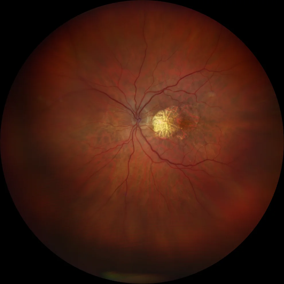
B1: geographic atrophy has extended to the temporal area after 4 years of follow-up. This new area of atrophy, however, is not as marked as the initial area.
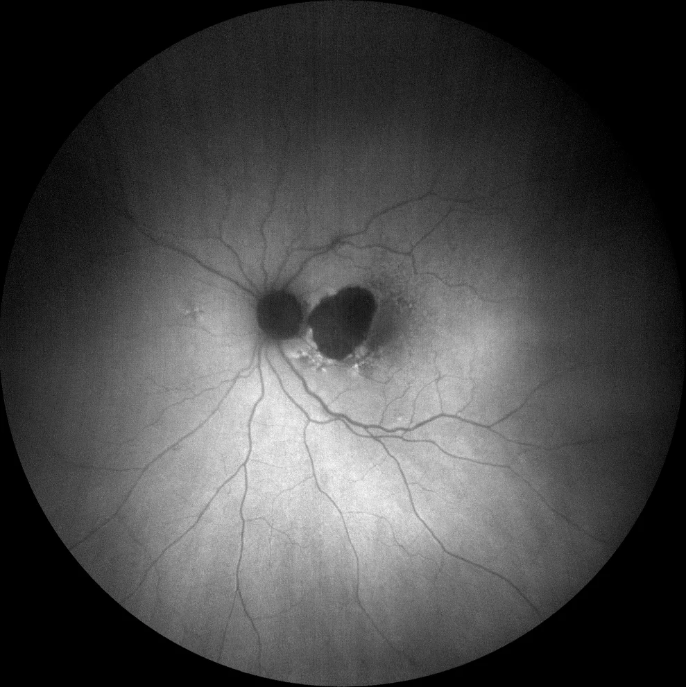
A2 (green AF): diffuse perilesional hyperAF pattern (reticular type)

B2 (green AF): the increase in the area of atrophy after 4 years of follow-up leaves a central island of healthy retina
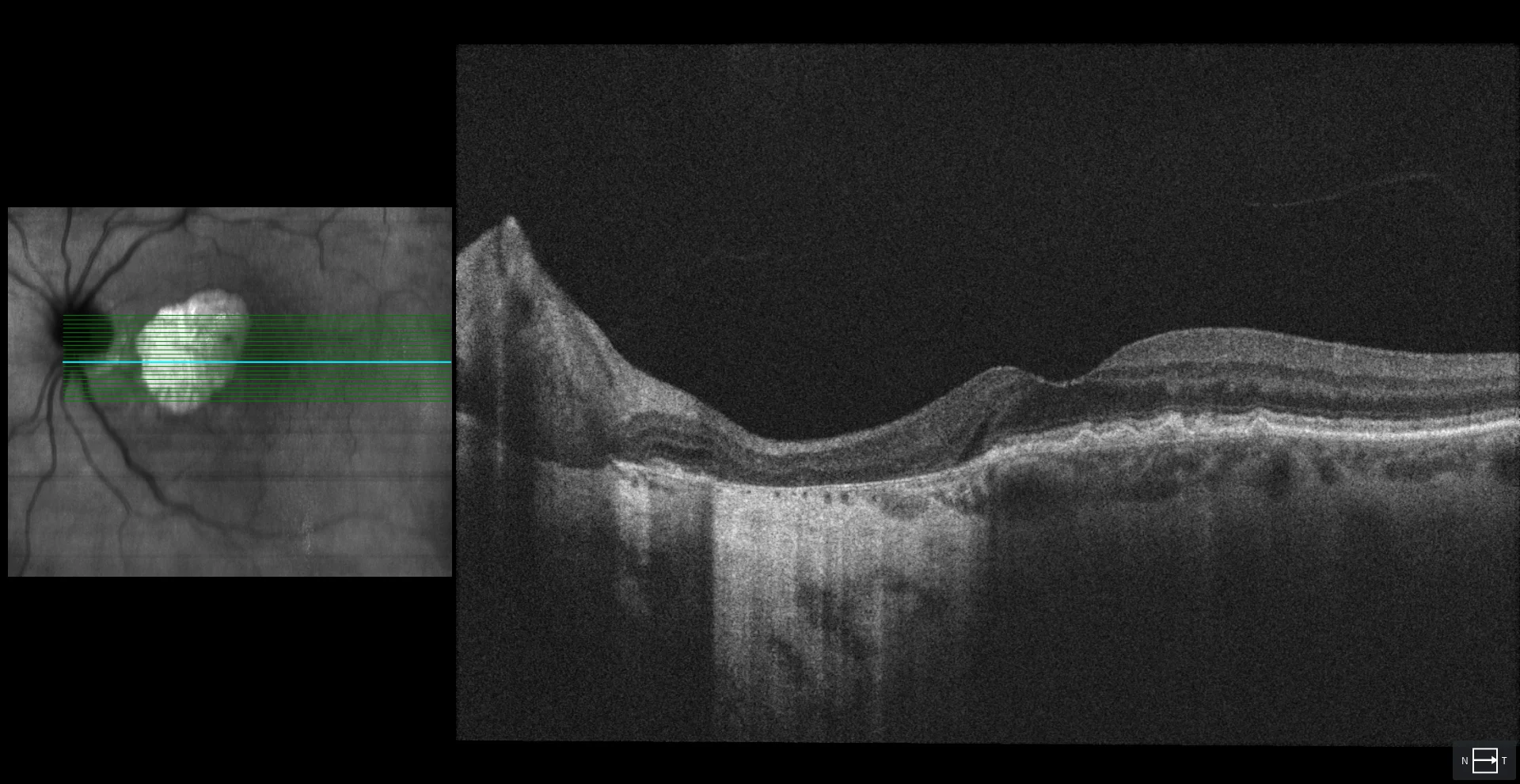
A3: In the foveal section, a cRORA (complete atrophy of the RPE and retina) is seen nasal to the fovea.
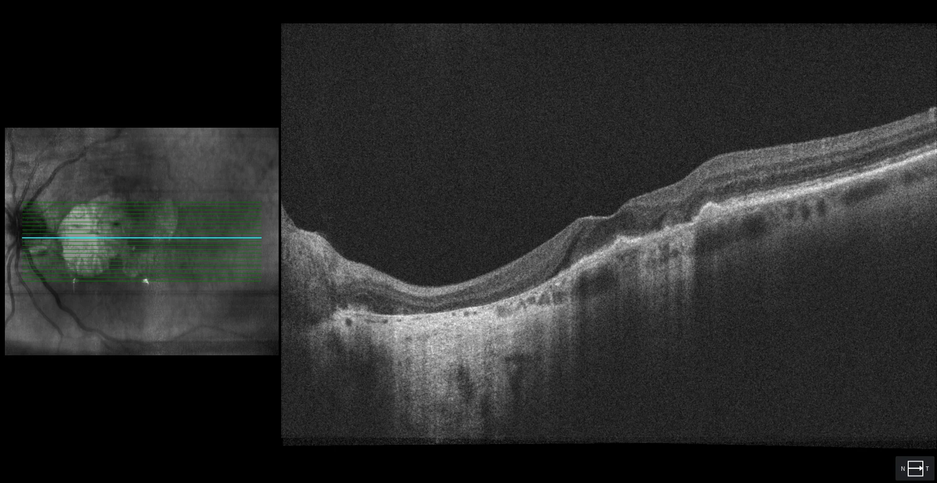
B3: temporal to the fovea, iRORA (incomplete RPE and retinal atrophy) is seen after 4 years of follow-up. As it is an area of incomplete atrophy, it is not seen as intensely as the cRORA area in the retinography (B1).
B4: OCT en face after 4 years of follow-up. Segmenting in choroid, the area of RPE atrophy is perfectly delimited.
Description
78-year-old woman who comes to the consultation for a check-up.
VA in LE is 20/20. An area of extrafoveal geographic atrophy is observed in the fundus, which is confirmed by autofluorescence and OCT (A).
After 4 years of follow-up, the VA in LE is 20/40. The area of geographic atrophy has extended, leaving a central island of healthy retina (B).