Maternally inherited diabetes and deafness (MIDD, Ballinger-Wallace syndrome)
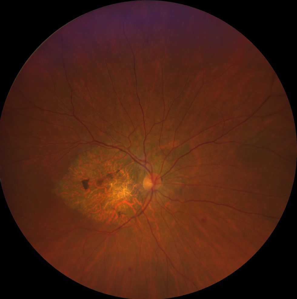
Color retinographies (Clarus 500, Carl Zeiss) of the right and left eyes of both sisters, showing an area of central atrophy in both eyes, more pronounced in the case of the older sister
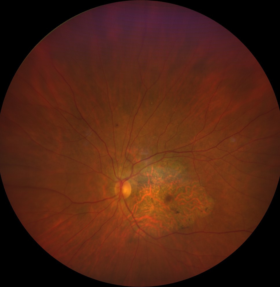
Color retinographies (Clarus 500, Carl Zeiss) of the right and left eyes of both sisters, showing an area of central atrophy in both eyes, more pronounced in the case of the older sister
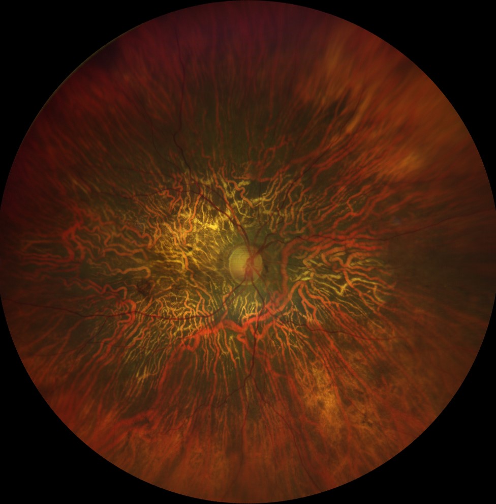
Color retinographies (Clarus 500, Carl Zeiss) of the right and left eyes of both sisters, showing an area of central atrophy in both eyes, more pronounced in the case of the older sister
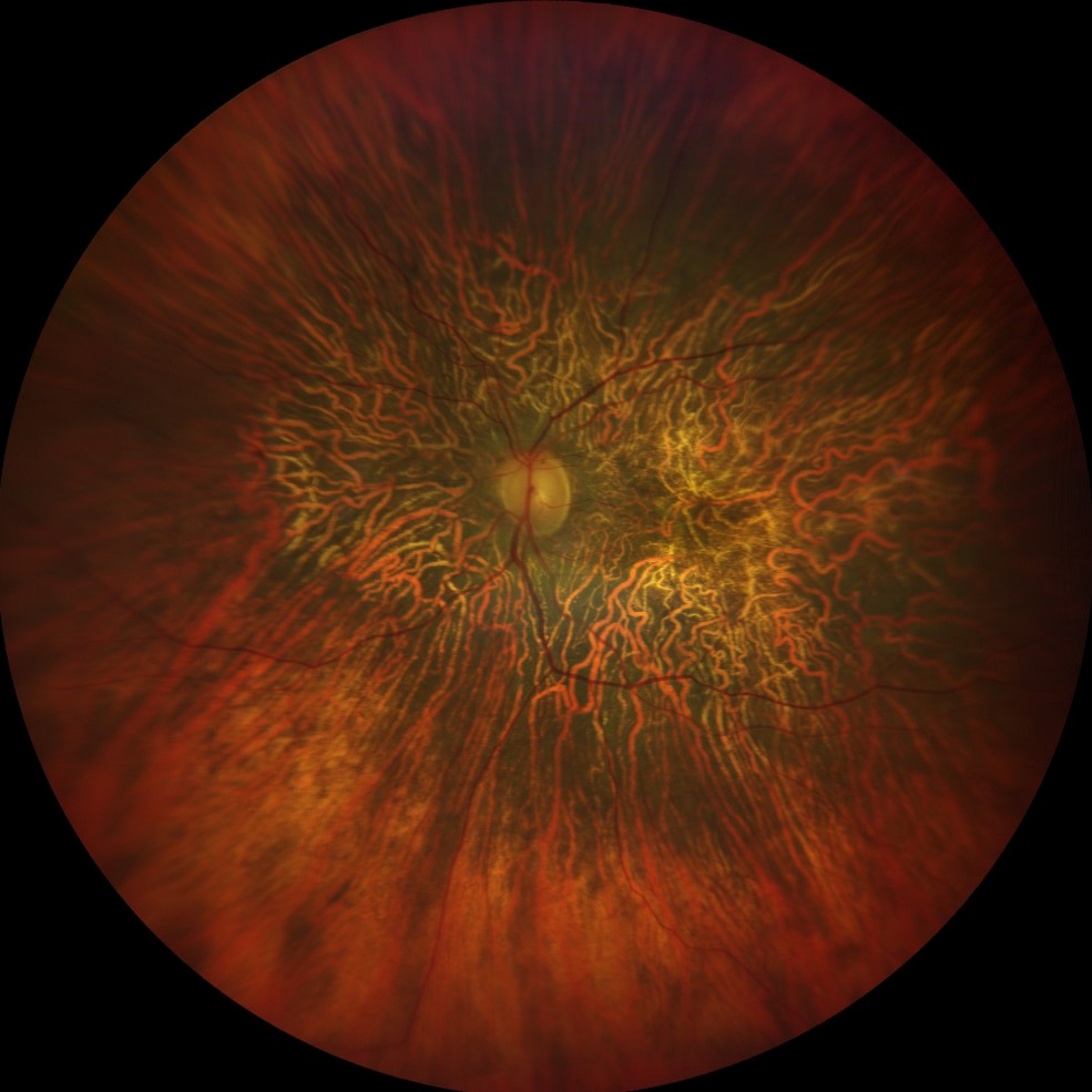
Color retinographies (Clarus 500, Carl Zeiss) of the right and left eyes of both sisters, showing an area of central atrophy in both eyes, more pronounced in the case of the older sister
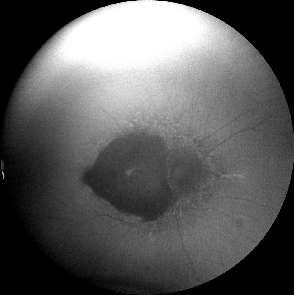
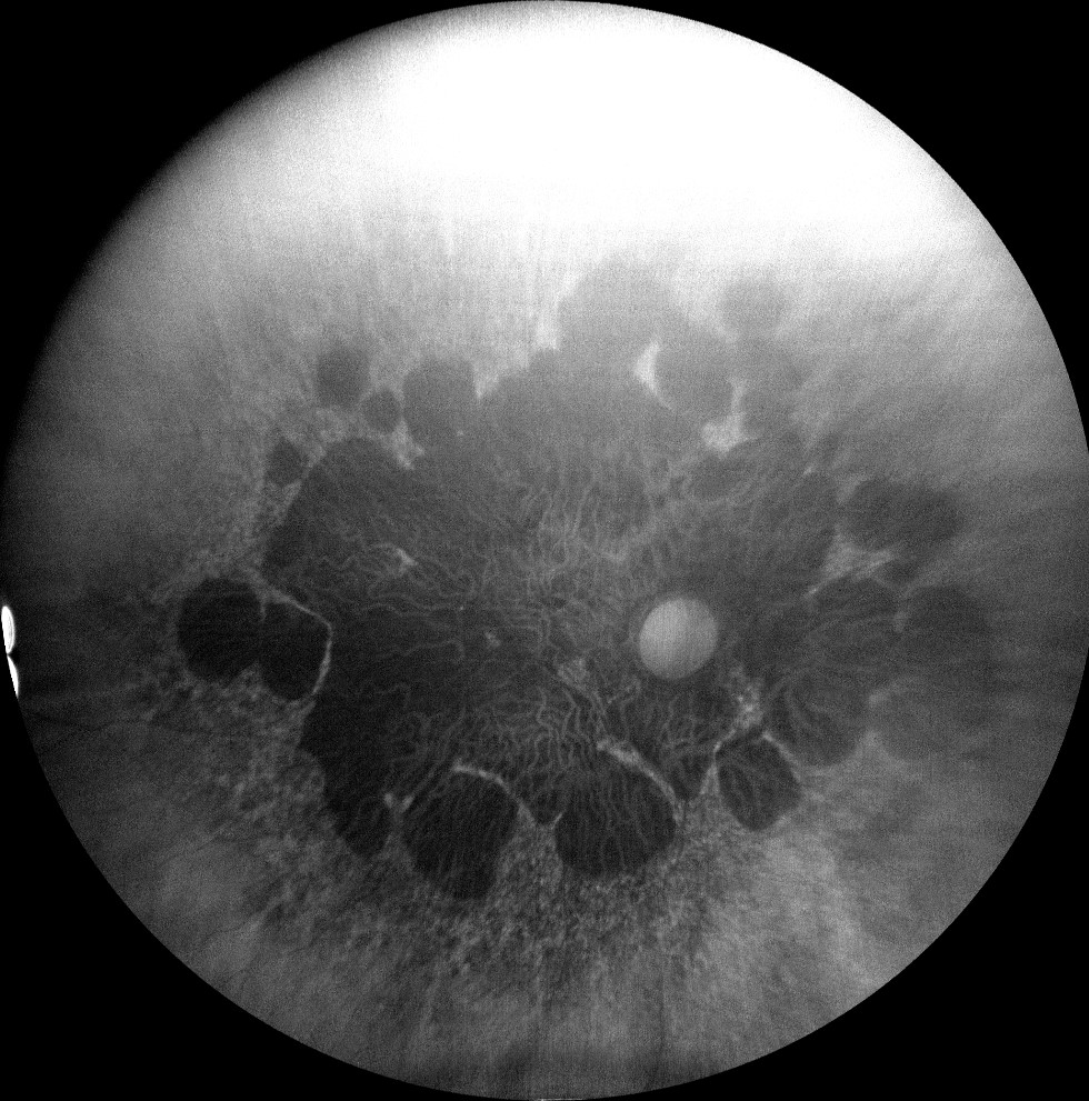
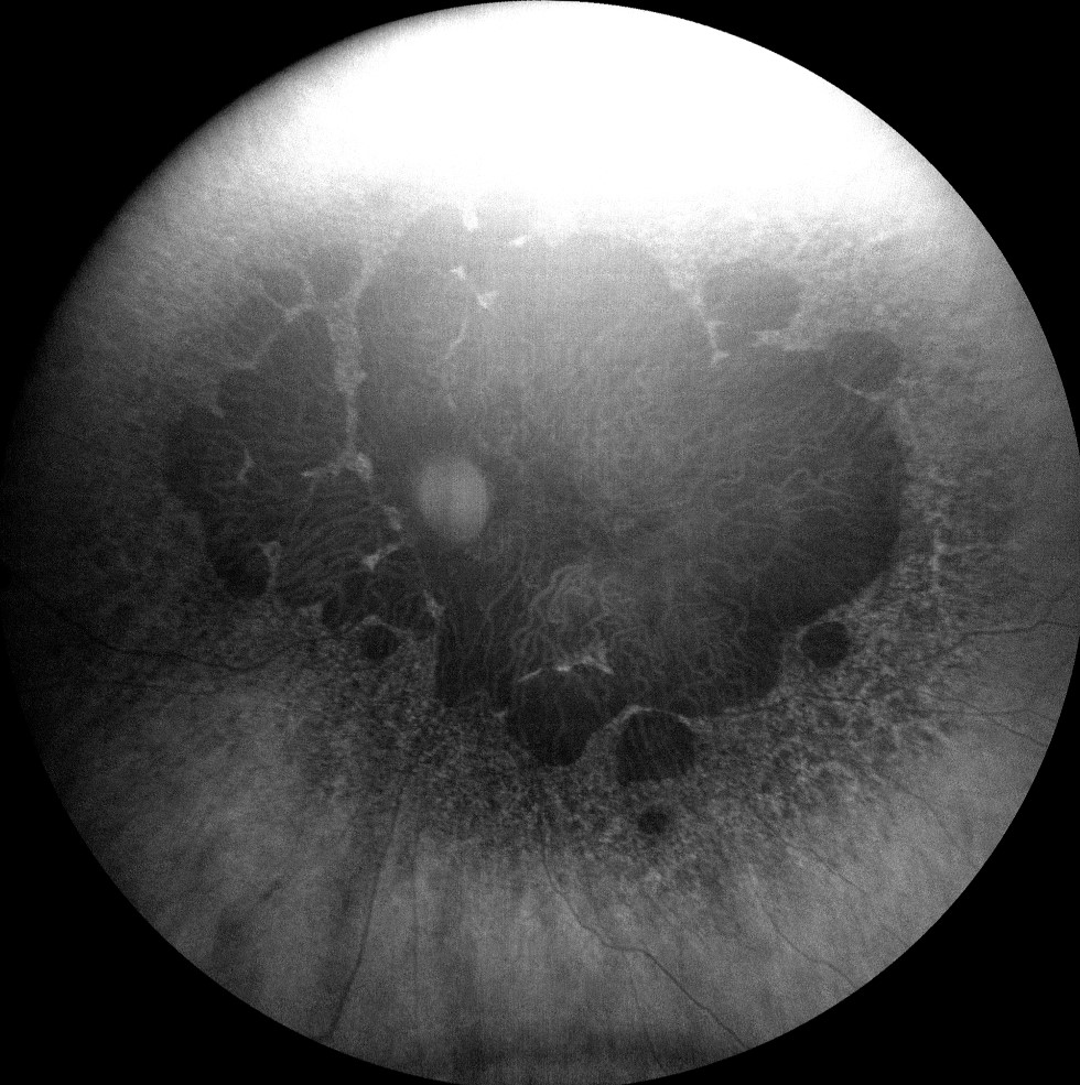
Autofluorescence images (Clarus 500, Carl Zeiss) of the right and left eyes of both sisters, showing hypoautofluorescence of atrophic macular lesions.
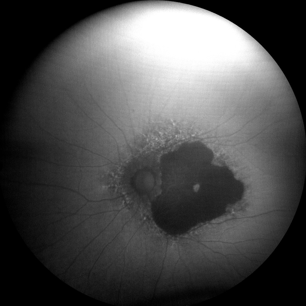
Autofluorescence images (Clarus 500, Carl Zeiss) of the right and left eyes of both sisters, showing hypoautofluorescence of atrophic macular lesions.

Autofluorescence images (Clarus 500, Carl Zeiss) of the right and left eyes of both sisters, showing hypoautofluorescence of atrophic macular lesions.
Autofluorescence images (Clarus 500, Carl Zeiss) of the right and left eyes of both sisters, showing hypoautofluorescence of atrophic macular lesions.
Autofluorescence images (Clarus 500, Carl Zeiss) of the right and left eyes of both sisters, showing hypoautofluorescence of atrophic macular lesions.
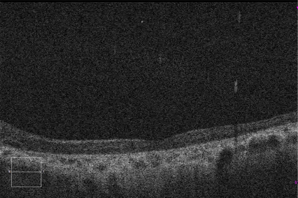
Macular HD optical coherence tomography (Cirrus 5000, Carl Zeiss) of the right and left eyes of both sisters, showing complete atrophy of the outer layers of the retina and the macular pigment epithelium.
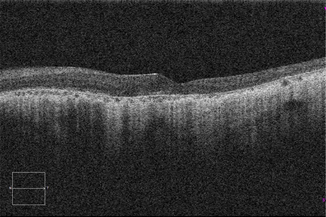
Macular HD optical coherence tomography (Cirrus 5000, Carl Zeiss) of the right and left eyes of both sisters, showing complete atrophy of the outer layers of the retina and the macular pigment epithelium.
Description
Maternally inherited diabetes and deafness (MIDD) is a very rare form of diabetes with a mitochondrial inheritance pattern caused by the 3243A mutation.> G of mitochondrial DNA. It is characterized by being associated with deafness even before the onset of diabetes and in up to 86% a macular atrophy is observed in a characteristically central areolar pattern.
Patients may report vision loss, night blindness, scotoma, or photophobia. These symptoms usually do not occur until there are significant alterations in the RPE. º